Department of Sonography

  • Home
  • Department of Sonography

Department overview

Sonography is the branch of Imaging Science through which human body organs can be scanned for any deviation from normalcy through high frequency sound waves.

Department of Sonography

Facilities

  • Per Abdomen Sonoraphy.
  • Transvaginal Sonogrphy.
  • Transrectal Sonography.
  • Soft Tissue Sonography

Gynaec Sonograpgy

  • Is mainly done to scan Uterus Ovaries & other Pelvic Organs to detect presence of any Tumour, Cancer, Extension & Spread of Cancer.
  • Sonography In Infertility Couple.
  • To see size and shape of Uterus & Ovaries.
  • To see the Endomteriosis or Ovarian Cyst.
  • Follicular monitoring: Serial study for size of follicle along with endometrial lining is done to estimate precise timing of Ovulation.
  • Sonography of testis to rule out varicocele.
  • Sonosalpingography toestimate patency of Fallopian Tubes.
  • Abdominal Sonography: To detect various abnormality of abdominal organs like Liver, Gall Bladder, Kidneys, Pancreas, Intestines. Any tumour.
  • Soft Tissue Sonography for Eye, Thyroid, Breast.

Obstetric Sonography

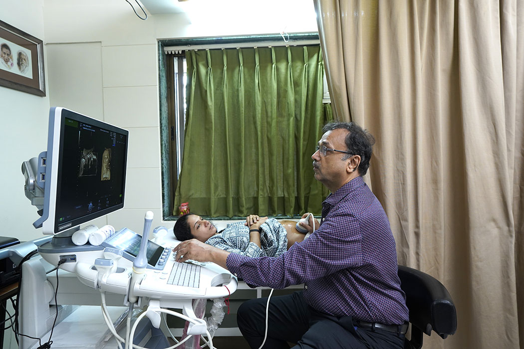
Obstetric Sonography is done for precise detection of fetal anomalies, proper growth of baby and for early detection of fetal health problems. It is done at various stages of pregnancy.

1st Trimester Scan (Upto 12 Weeks)

  • Diagnosis & location of pregnancy.
  • For number of fetus.
  • For nuchal translucency.
  • For early detection of certain anomalies.
  • For viability of fetus.
  • Placental location.

2nd Trimester Target Scan (At 20 weeks)

  • Each and every part of fetus is closely scanned for detection of anomalies of smallest part of fetus.
  • Fetal echocardiography to detect structural anomalies of fetal heart.
  • Fetoplacental perfusion in high risk pregnancy.
  • Amount of Liquor.

Scan at 32 weeks

  • To rule out fetal growth retardation.
  • For presentation.
  • For amount of Liquor.

Scan At Term (Near Due Date)

  • For Estimation of Fetal Weight
  • Fetal Presentation
  • Position of Placenta
  • Amount of Liquor
  • Color Doppler Sonography

Speciality

  • Color Doppler Sonography: It is done mainly to assess the perfusion of particular organ.
  • It is of great help to decide perfect time for IUI in ovulation monitoring, to increase success rate of IVF cycle.
  • It provides knowledge of proper feto placental circulation especially at term of pregnancy.
  • For detection of cardiac anomalies.
  • For diagnosis of varicocele in male infertility.
  • For peripheral doppler study of arteries veins of limbs.
  • Fetal Echocardiography.
  • Usually fetal heart evaluation is performed at 18-22 wks of pregnancy.
  • More than 50% of the cardiac anomalies can be corrected after birth, if they are timely diagnosed during antenatal sonography.
  • Presence of two or more extra cardiac anomalies increase the risk of cardiac anomaly up to 75%.
  • 20% of Non immune hydrops is associated with structural heart diseases.
  • Family history of Congenital heart diseases and maternal metabolic diseases like diabetes increases the risk of fetal cardiac anomaly.
  • 4D Ultrasonography: Done in NNH on Voluson Machine.
  • It is for acquisition of fetal face within Uterus.
  • Fetus seen with a smile an yawn. Thumb sucking, stretching, waving a hand is an exciting site to watch for would be parents and family members.
  • 3D Sonography is useful in precise detection of anomalous fetal spine.
  • Different anomalies of Uterus : Useful in Female infertility management.
  • For extent of Tumour.